297 results

- Digital Images
- Online
Confocal micrograph of E. coli
Fernan Federici & Jim Haseloff
- Digital Images
- Online
Confocal micrograph of E. coli
Fernan Federici & Jim Haseloff
- Digital Images
- Online
Confocal micrograph of E. coli
Fernan Federici & Jim Haseloff
- Digital Images
- Online
Confocal micrograph of Bacillus subtilis. Bacillus subtilis is a Gram-positive, rod-shaped bacterium, commonly found in soil. Fluorescent proteins (TagRFP-T, sfGFP, TagBFP, mKate2 and mOrange2), time-lapse confocal microscopy and biophysical models are being used to understand the organization of bacterial biofilms.
Fernan Federici & Jim Haseloff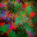
- Digital Images
- Online
Confocal micrograph of Bacillus subtilis. Bacillus subtilis is a Gram-positive, rod-shaped bacterium, commonly found in soil. Fluorescent proteins (TagRFP-T, sfGFP, TagBFP, mKate2 and mOrange2), time-lapse confocal microscopy and biophysical models are being used to understand the organization of bacterial biofilms.
Fernan Federici & Jim Haseloff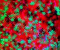
- Digital Images
- Online
Confocal micrograph of Bacillus subtilis. Bacillus subtilis is a Gram-positive, rod-shaped bacterium, commonly found in soil. Fluorescent proteins (TagRFP-T, sfGFP, TagBFP, mKate2 and mOrange2), time-lapse confocal microscopy and biophysical models are being used to understand the organization of bacterial biofilms.
Fernan Federici & Jim Haseloff
- Digital Images
- Online
Hippocampal neurone, confocal image
Claudius Griesinger
- Digital Images
- Online
Hippocampal neurone, confocal image
Claudius Griesinger
- Digital Images
- Online
Neuroblast spindle, confocal image
Dr Andrea H. Brand
- Digital Images
- Online
Segregating determinants in embryo, confocal
Dr Andrea H. Brand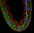
- Digital Images
- Online
Neural precursors in embryo, confocal
Dr Andrea H. Brand
- Digital Images
- Online
Pioneering neurons in embryo, confocal image
Dr Andrea H. Brand
- Digital Images
- Online
Neuroblast cell division in embryo, confocal
Dr Andrea H. Brand
- Digital Images
- Online
Axons in developing embryo, confocal image
Dr Andrea H. Brand
- Digital Images
- Online
Axons in living embryo, confocal image.
Dr Andrea H. Brand
- Digital Images
- Online
Mouse embryonic posterior neuropore, confocal image.
Galea, Gabriel.Date: 2016
- Digital Images
- Online
Neuroblasts in developing embryo, confocal image
Dr Andrea H. Brand
- Digital Images
- Online
Interneuron, stereo confocal image
Biosciences Imaging Gp, Soton