14 results

- Pictures
- Online
The muscles of the hand, after Albinus, and of the foot, after De Courcelles. Engraving by Benard, late 18th century.
Wandelaar, Jan, 1690 or 1692-1759.Reference: 35337i
- Pictures
- Online
The muscles of the hand, after Albinus, and of the foot, after De Courcelles. Engraving by Benard, late 18th century.
Wandelaar, Jan, 1690 or 1692-1759.Reference: 35352i
- Pictures
- Online
The muscles of the human body, first layer, seen from the front, after Albinus. Engraving by Benard, late 18th century.
Wandelaar, Jan, 1690 or 1692-1759.Reference: 35293i
- Pictures
- Online
The muscles of the human body, first layer, seen from the back, after Albinus. Engraving by Benard, late 18th century.
Wandelaar, Jan, 1690 or 1692-1759.Reference: 35349i
- Pictures
- Online
The muscles of the human body, first layer, seen from the back, after Albinus. Engraving by Benard, 1779, after an engraving, 1747.
Wandelaar, Jan, 1690 or 1692-1759.Date: [1779]Reference: 36173i
- Pictures
- Online
The muscles of the human body, first layer, seen from the front, after Albinus. Engraving by Benard, 1779, after an engraving, 1747.
Wandelaar, Jan, 1690 or 1692-1759.Date: [1779]Reference: 36172i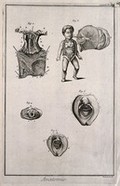
- Pictures
- Online
The uterus, a foetus, the hymen and female genitals, after Haller, Kulm and Huber. Engraving by Benard, late 18th century.
Reference: 35218i
- Pictures
- Online
The arteries of the thorax, after Haller. Engraving by Benard, late 18th century.
Kaltenhofer, Joel Paul, -1777.Reference: 35065i
- Pictures
- Online
The female generative organs, after Haller. Engraving by Benard, late 18th century.
Rollinus, C. J., active 18th century.Reference: 35209i
- Pictures
- Online
Nervous system after Vieussens (fig. 1); brain and spinal cord after Eustachius (fig. 2) Engraving by Benard, late 18th century.
Reference: 34440i
- Pictures
- Online
The viscera, after Haller, showing the lobes of the liver, the stomach, the kidneys, the spleen, etc. Engraving by Benard, late 18th century.
Rollinus, C. J., active 18th century.Reference: 35068i
- Pictures
- Online
The arteries of the head after Haller; the eye, after Ruysch, Cowper and Bidloo. Engraving by Benard, late 18th century.
Rollinus, C. J., active 18th century.Reference: 34482i
- Pictures
- Online
The arteries of the head after Haller; the eye, after Ruysch; the tongue after Heister. Engraving by Benard, late 18th century.
Rollinus, C. J., active 18th century.Reference: 34490i
- Pictures
- Online
The diaphragm (fig. 1) after Haller, the pharynx, seen from the back and the larynx seen from the front (figs 2-3), after Duverney, and the larynx seen from the back and open and from the side (figs 4-5), after Eustachius. Engraving by Benard, late 18th century.
Reference: 35356i