66 results
- Pictures
- Online
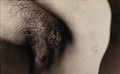
An armpit (with a growth ?). Photograph, ca. 1890.
Date: 1890Reference: 571895i
- Pictures
- Online
An operation in progress: an incision in the armpit. Photograph by Félix Méheux, ca. 1900.
Méheux, Félix.Date: 1900Reference: 570584i- Books
Why the watermelon won't ripen in your armpit : and other science conundrums / Ben Selinger.
Selinger, B. K. (Benjamin Klaus)Date: 2000
- Pictures
- Online
An operation in progress: a metal clip in an incision in the armpit. Photograph by Félix Méheux, ca. 1900.
Méheux, Félix.Date: 1900Reference: 570592i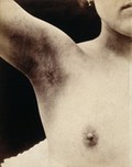
- Pictures
- Online
A breast operation to remove a lump, in progress: scarring to the armpit following the operation. Photograph by Félix Méheux, ca. 1900.
Méheux, Félix.Date: 1900Reference: 570619i
- Pictures
- Online
An operation in progress: a metal surgical implement is placed inside an incision in the armpit. Photograph by Félix Méheux, ca. 1900.
Méheux, Félix.Date: 1900Reference: 570587i
- Pictures
- Online
An operation in progress: a metal surgical implement is placed inside an incision in the armpit. Photograph by Félix Méheux, ca. 1900.
Méheux, Félix.Date: 1900Reference: 570591i- Pictures
A Chinese man displaying symptoms of axillary anthrax, in the armpit area. Coloured line block print by Chiang Yee, after a Chinese artist, 1920/1940?.
Date: [1920/1940?]Reference: 573725i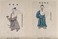
- Pictures
- Online
A Chinese man displaying symptoms of an axillary carbuncle on his armpit. Coloured line block print by Chiang Yee, after a Chinese artist, 1920/1940?.
Date: [1920/1940?]Reference: 573478i
- Pictures
- Online
The circulatory system: dissection of the upper arm, shoulder and armpit, with arteries and blood vessels indicated in red. Coloured lithograph by J. Maclise, 1841/1844.
Maclise, Joseph.Date: [1841/1844]Reference: 579567i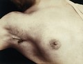
- Pictures
- Online
A breast operation to remove a lump, in progress: an incision in the armpit which has been sewn up. Photograph by Félix Méheux, ca. 1900.
Méheux, Félix.Date: 1900Reference: 570617i- Pictures
The upper humerus bone and socket, highlighting a bone marrow tumor in the armpit in a 42-year old woman with leukaemia. Radiographic print, 1946.
Nicholson, BarbaraDate: 1946Reference: 31317iPart of: Barbara Nicholson medical illustration collection.- Pictures
Cystic hygroma of armpit in a twenty year old woman with swelling and lump: surgical specimen detail shown from above. Watercolour by Barbara E. Nicholson, 1951.
Nicholson, BarbaraDate: 1951Reference: 34271iPart of: Barbara Nicholson medical illustration collection.- Pictures
Areas of diseased skin around the armpit and genitals of a man suffering from tinea circinata (ringworm of the body): two figures. Chromolithograph by E. Burgess, 1850/1880?.
Burgess, E.Date: [1850/1880?]Reference: 578104i
- Pictures
- Online
A breast operation to remove a lump, in progress: a metal surgical implement is placed inside an incision in the armpit. Photograph by Félix Méheux, ca. 1900.
Méheux, Félix.Date: 1900Reference: 570595i- Pictures
Cystic hygroma of armpit in a twenty year old woman with swelling and lump: surgical specimen detail showing dilated lymphatic cavity. Watercolour by Barbara E. Nicholson, 1951.
Nicholson, BarbaraDate: 1951Reference: 34206iPart of: Barbara Nicholson medical illustration collection.
- Pictures
- Online
The circulatory system: dissection of the upper arm, shoulder and armpit, with arteries, blood vessels and veins indicated in red and blue. Coloured lithograph by J. Maclise, 1841/1844.
Maclise, Joseph.Date: [1841/1844]Reference: 579570i
- Pictures
- Online
A breast operation to remove a lump, in progress: an incision in the armpit; the breast is pinched between thumb and forefinger. Photograph by Félix Méheux, ca. 1900.
Méheux, Félix.Date: 1900Reference: 570594i
- Pictures
- Online
A surgical operation to remove a malignant tumour from a man's left breast and armpit in a Dublin drawing room, 1817. Watercolour, ca 1913, after a watercolour, 1817.
Date: 1913Reference: 23451i
- Pictures
- Online
A breast operation to remove a lump, in progress: a metal surgical implement is placed into the breast via an incision in the armpit. Photograph by Félix Méheux, ca. 1900.
Méheux, Félix.Date: 1900Reference: 570599i
- Pictures
- Online
A breast operation to remove a lump, in progress: metal surgical tongs hold a lump taken from the breast via an incision in the armpit. Photograph by Félix Méheux, ca. 1900.
Méheux, Félix.Date: 1900Reference: 570606i- Pictures
The upper humerus bone and socket, highlighting a bone marrow tumor in the armpit in a 42-year old woman with leukaemia. Pen and ink drawing by Barbara E. Nicholson after a radiograph, 1946.
Nicholson, BarbaraDate: 1946Reference: 31831iPart of: Barbara Nicholson medical illustration collection.
- Pictures
- Online
The circulatory system: dissection of the armpit of a man, showing the musculature and lymph nodes and with arteries, blood vessels and veins indicated in red and blue. Coloured lithograph by J. Maclise, 1841/1844.
Maclise, Joseph.Date: [1841/1844]Reference: 579574i
- Pictures
- Online
Left, a man suffers the "torture of water"; left, a man tied to a rack has his armpits and the soles of his feet scorched by candles. Etching.
Reference: 43400i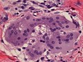
- Digital Images
- Online
Multinucleated giant cell containing an asteroid, microscopy.
William R. Geddie