402 results filtered with: Digital Images, Pictures

- Digital Images
- Online
3D reconstruction of chick heart
NIMR, MRC
- Digital Images
- Online
3D printed bioglass lattice, SEM
Ezra Feilden
- Digital Images
- Online
3D reconstruction of chinchilla, composite
Scott Birch
- Digital Images
- Online
3D reconstruction of chinchilla
Scott Birch
- Digital Images
- Online
3D reconstruction of chinchilla
Scott Birch
- Digital Images
- Online
3D modelling of proteins on computer
Mol. Biophysics, Oxford Univ.
- Digital Images
- Online
3D modelling of proteins on computer
Mol. Biophysics, Oxford Univ.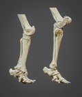
- Digital Images
- Online
3D reconstructed elephant hind limbs
Scott Birch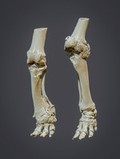
- Digital Images
- Online
3D reconstructed elephant hind limbs
Scott Birch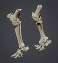
- Digital Images
- Online
3D reconstructed elephant hind limbs
Scott Birch
- Digital Images
- Online
3D reconstructed elephant hind limbs
Scott Birch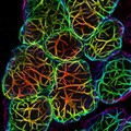
- Digital Images
- Online
3D depth-coloured transparent mouse mammary gland
Felicity Davis, Bethan Lloyd-Lewis and Christine Watson; University of Cambridge
- Digital Images
- Online
3D-printed reconstruction of a healthy adult human brain
Stephanie J. Forkel, Natbrainlab, King's College London, Ahmad Beyh, Natbrainlab, King's College London & Michel Thiebaut de Schotten, Brain Connectivity and Behaviour Group, Brain and Spine Institute, La Salpêtrière
- Digital Images
- Online
3D printed reconstruction of the arcuate fasciculus.
Forke, Stephanie J.Date: 2016
- Digital Images
- Online
3D MRI of a 6-day-old quail egg
Suzanne Duce / University of Dundee
- Digital Images
- Online
3D print of vessels of a healthy mini pig eye.
Maloca, Peter M.Date: 2016
- Digital Images
- Online
3D MRI of a six-day-old quail embryo alive inside its egg.
Suzanne Duce / University of Dundee
- Digital Images
- Online
3D MRI of a six-day-old quail embryo alive inside its egg.
Suzanne Duce / University of Dundee
- Digital Images
- Online
3D MRI of a six-day-old quail embryo alive inside its egg
Suzanne Duce / University of Dundee
- Digital Images
- Online
3D view on vessels of a healthy minipig eye. The upper opening corresponds to the pupil as the gateway input of all light into the eye. It is interesting to see the marked abundance of vessels of the pupil which bring energy and food to the muscles to control the amount of incident light. The other large vessels are feeder vessels for the outer layers of the retina and muscles, so, that the eye quickly can perceive the environment and the creature may adapt and survive.
Peter M Maloca, Christian Schwaller, Ruslan Hlushchuk, Sébastien Barré, OCTlab University of Basel/Bern and Royal Moorfields Eye Hospital, London
- Digital Images
- Online
Freshwater diatoms SEM 3D image
Biosciences Imaging Gp, Soton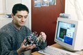
- Digital Images
- Online
Researcher with 3-D model of protein
Mol. Biophysics, Oxford Univ.
- Digital Images
- Online
Researcher with 3-D model of protein
Mol. Biophysics, Oxford Univ.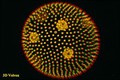
- Digital Images
- Online
Volvox (colonial freshwater alga) SEM, 3D
Biosciences Imaging Gp, Soton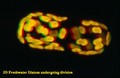
- Digital Images
- Online
Freshwater diatom dividing, SEM, 3D image
Biosciences Imaging Gp, Soton