Licence: In copyright
Credit: On Eurycarpus oweni / H.G. Seeley. Source: Wellcome Collection.
Provider: This material has been provided by The Royal College of Surgeons of England. The original may be consulted at The Royal College of Surgeons of England.
7/12 page 329
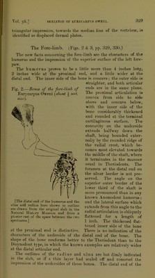
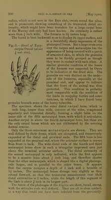
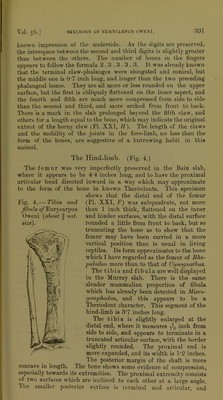
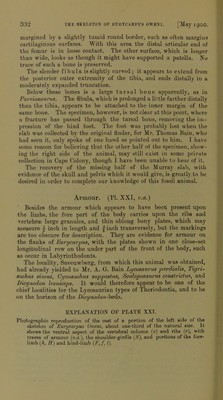
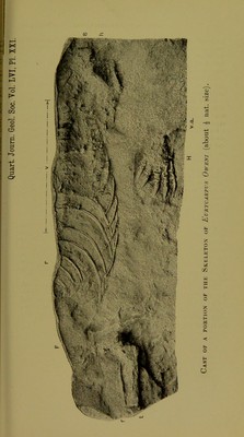
![triangular impression, towards the median line of the vertcbrte, is identified as displaced dermal plates. asek The Fore-lirab. (Figs. 2 & 3, pp. 329, 330.) The new facts concerning the fore-limb are the characters of the humerus and the impressiou of the superior surface of the left fore- paw. The humerus proves to be a little more than 4 inches long, 2 inches wide at the proximal end, and a little wider at the distal end. The inner side of the bone is concave; the outer side is straighter, and both articular Fig. 2.—Bones of the fore-limb of ends are in the same plane. Eurycarpus Oweni (about ^ not. proximal articulation is size\ convex from side to side above and concave below, with the inner side of the bone considerably thickened and rounded at the terminal cartilaginous surface. The concavity on the underside extends halfway down the shaft, being bounded exter- nally by the rounded ridge of the radial crest, which be- comes most elevated towards the middle of the shaft, where it terminates in the manner usual in Theriodonts. The foramen at the distal end on the ulnar border is not pre- served. The angle on the superior outer border of the lower third of the shaft is more pronounced than in any known Anomodont humerus; and the lateral surface which extends from this angle to the radial articulation is obliquely flattened for a length of 1 inch. The thickened flat- tened inner side of the bone at the proximal end is distinctive. There is no indication of the characters of the underside of the distal end of the bone. The shape of the bone conforms better to the Theriodont than to the Dicynodont type, in which the known examples are relatively wider at the distal articular end. Ihe outlines ot the radius and ulna are but dimly indicated in the slab, as if a thin layer had scaled off and removed the impression of the undersides of those bones. The distal end of the [The distal end of t.he b umerim and the ulna and radius here shown in outline are drawn from the original slab in the Natural History Museum and from a plaster east of the space between the two specimens.]](https://iiif.wellcomecollection.org/image/b22412888_0009.jp2/full/800%2C/0/default.jpg)
 No text description is available for this image
 No text description is available for this image
 No text description is available for this image