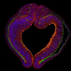
Confocal micrograph showing an optical section through the optic vesicle (early eye) in a chick. At this stage of development the chick embryo has 8 somites which is approximately 36 hours after the egg was laid. Cells in the optic vesicle have been stained with antibodies against different proteins to visualise them. The cell membrane of individual epithelial cells (red; beta-catenin) and their nuclei (blue; DAPI) are visible. Below the epithelial cells sits the basal lamina (green; laminin) which provides support to the overlying sheets of cells as well as allowing small molecules and water to pass through. The protein neural cadherin (N-cadherin) is shown in yellow and labels the apex of the epithelial cells (stained in red). Cadherins play an important role in cell adhesion helping bind cells within a tissue together. The chick is used as a model system to study development of the eye in order to better understand how defects develop in certain conditions. Congenital anophthalmia and microphthalmia (AM) are developmental defects resulting in the absence or reduction of the eye, with a combined incidence of up to 30 per 100,000 births. In most cases, the cause is poorly understood. Horizontal width of image is approximately 800 microns.