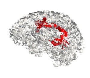
Visualising language processing regions in a living human brain. In this image the brain is viewed from the side (sagittal view), with the front of the brain facing the left side of the image and the back of the brain on the right. Language is processed across a large network of brain regions, with two regions primarily important for language, one for articulation and one for comprehension. These areas are located in distant parts of the brain and are connected to each other by the arcuate fasciculus (red).
Brain cells communicate with each other through these nerve fibres, which have been visualised using diffusion imaging tractography. Diffusion weighted imaging is a specialised type of magnetic resonance imaging (MRI) scan which measures water diffusion in many directions in order to reconstruct the orientation of bundles of axons. Tractography is used to indirectly model these bundles of axons (nerve fibres), which transmit information between cortical regions at the brain's surface.
These connections are being studied in the healthy brain in order to further understand changes that occur in developmental disorders (for example autism), neurodegenerative processes (such as dementia) and head trauma (for example stroke).