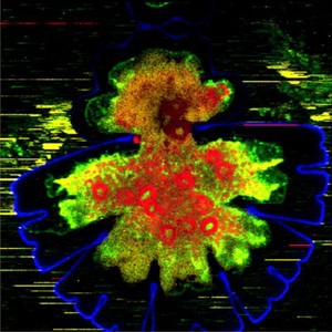
Confocal Raman micrograph of the desmid Micrasterias denticulata undergoing cell division. Micrasterias is a unicellular green alga mostly found in neutral to acidic fresh waters and sphagnum bogs. Raman spectroscopy is a technique used to collect information about the chemical fingerprint of molecules. Here, this information is being visualised by combining it with confocal microscopy. The outer cell wall (blue), starch (red), and proteins, pectins and fats (green and yellow) are visible. The cell wall is more highlighted in the older half of the cell (lower part of image, larger in size) due to higher amounts of cellulose and crystallinity than in the newly formed younger part (upper part of image, smaller in size). Inside the algae, starch and small round pyrenoids (red) embedded in the chloroplast are visible in the older half of the cell. Width of image is 160 micrometres.