HIV integration, HIV viral life cycle, illustration

  • David S. Goodsell, The Scripps Research Institute
  • Digital Images
  • Online

Available online

view HIV integration, HIV viral life cycle, illustration

Attribution 4.0 International (CC BY 4.0)

You can use this work for any purpose, including commercial uses, without restriction under copyright law. You should also provide attribution to the original work, source and licence. Read more about this licence.

Credit

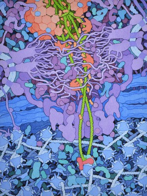
HIV integration, HIV viral life cycle, illustration. David S. Goodsell, The Scripps Research Institute. Attribution 4.0 International (CC BY 4.0). Source: Wellcome Collection.

Selected images from this work

View 1 image

About this work

Description

Watercolour and ink on paper illustration of a cross section through part of a target cell (blue and purple; usually a CD4 T cell, a type of white blood cell found in the immune system) infected with HIV. Once the virus has entered the target cell, the viral capsid uncoats (red; top middle) and interacts with nuclear pore proteins such as Nup358 releasing the viral DNA (green). Viral DNA enters the nucleus (lower part of image) from the cytoplasm (upper part of image) through the nuclear pore (purple) and is integrated into the cell's own genome by the enzyme HIV integrase (red molecule; lower middle). Width of image is approximately 100 nm. This image forms part of a series of illustrations which capture HIV at eight different points in the viral life cycle as it infects a cell and replicates.

Permanent link