Pregnant uterus, Red deer

  • Michael Frank, Royal Veterinary College
  • Digital Images
  • Online

Available online

view Pregnant uterus, Red deer

Attribution-NonCommercial 4.0 International (CC BY-NC 4.0)

You can use this work for any purpose, as long as it is not primarily intended for or directed to commercial advantage or monetary compensation. You should also provide attribution to the original work, source and licence. Read more about this licence.

Credit

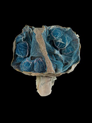
Pregnant uterus, Red deer. Michael Frank, Royal Veterinary College. Attribution-NonCommercial 4.0 International (CC BY-NC 4.0). Source: Wellcome Collection.

Selected images from this work

View 1 image

About this work

Description

Photograph of a pregnant red deer (Cervus elaphus) uterus with the fetus removed. The cut umbilical cord can be seen. The blood vessels have been injected with blue and red latex to make them more visible. Cotyledons (large spherical structures; blue) are the location where maternal and fetal circulation comes into contact so that nutrients can be exchanged.

Permanent link