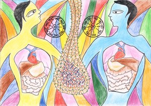
The picture depicts the biology of a very rare, but fascinating disease called situs inversus, first described in the late 17th century, where the visceral organs (such as the heart, lungs and digestive organs) are all laterally inverted within the body. This occurrence is due to dysfunction of a unique organelle, the motile cilia, in an embryonic cavity called the node that develops at the tip of the embryonic axis. The man on the left (yellow) depicts an individual with normal situs, whereas the one on the right (blue) depicts an individual afflicted with situs inversus. The node - which appears like a tear drop-shaped pit, and the notochord - which the node extends into anteriorly, are shown in the middle of the image. The cells within the pit of the node have motile cilia (blue), whereas the rest of the cells have immotile primary cilia (green).
Cilia are microtubule based finger like projections from cells. They can be motile and generate fluid flow, or immotile and serve as antennae for sensing inter-cellular signals. Within the node pit, the motile cilia generate a directed fluid flow sensed by the immotile primary cilia on the cells surrounding the pit. This then triggers a signalling cascade that initiates asymmetric morphogenesis of organs.
Motile cilia can beat rhythmically due to the presence of arms of dynein (a 'motor' protein) on their outer microtubule doublets (cross-sectional diagram of normal motile cilia structure on the left). The majority of cases of situs inversus arise from defects in these dynein arms, which render the cilia dysmotile or completely immotile (diagram on the right showing absence of dynein arms). Situs inversus is not dangerous by itself; people with this disorder lead a completely normal life. However, there are individuals in which the organ system reversal is not complete: a condition called heterotaxy. This often leads to severe complications, like congenital heart defects, which require corrective surgery.