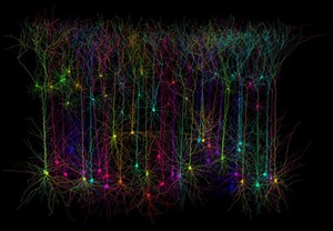
This computer simulated image was created using software called the 'TREES toolbox', which allows scientists to generate neuronal structures indistinguishable from those found in the real biological brain. This image shows synthetic neurons that represent the optimised size, shape and connectivity of pyramidal neurons analogous to those found in the cortex of the brain. Pyramidal neurons are so-called because they have a pyramid-shaped cell body (soma), they are also characterised by long branching dendrites. They are found in the forebrain (cortex and hippocampus) of mammals and are thought to be involved in cognitive function.
Each neuron is assigned a different colour so that individual structures and processes can be easily distinguished. These highly accurate synthetic neurons have the added advantage of being manipulated and viewed in a number of different ways, helping scientists learn more about cell shape and how neurons are constructed.
2011 Wellcome Image Award winner. Wellcome Image Awards 2011.