Volume 1
A system of anatomical plates of the human body; accompanied with descriptions, and physiological, pathological & surgical observations. Text and plates / By John Lizars.
- John Lizars
- Date:
- [1840?]
Licence: Public Domain Mark
Credit: A system of anatomical plates of the human body; accompanied with descriptions, and physiological, pathological & surgical observations. Text and plates / By John Lizars. Source: Wellcome Collection.
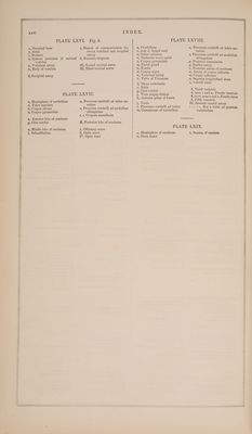
520/535 page 29
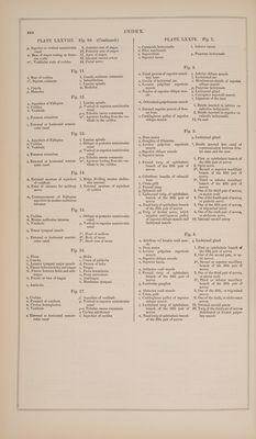
![a, Fallopian aqueduct w, Zygomatic process of temporal bone z, Osseous portion of Eustachian tube m*, Mastoid cells o, Foramen ovale p, Osseous pyramid of tympanum r, Foramen rotundum t, Tympanic cavity Fig. 6. a, Promontory of tympanum c, Protuberance made by external semicircular canal 1**, Bristle indicating semi-osse- ous canal of tensor tympani muscle 2, Bristle indicating Fallopian aqueduct, and emergence of chorda tympani nerve c, Cochlea M, Inferior recess of meatus audi- torius internus, which gives entrance to auditory nerve v, Vestibule z, Osseous portion of Eustachian tube m, Commencement of Fallopian aqueduct m*, Mastoid cells b, Ridge dividing meatus audito- rius Internus c, External or horizontal semicir- cular canal 0, Oblique or posterior semicircular canal canal munis 1*, Head of malleus 3, Long slender process of malleus 30, Handle of malleus 6*, Body of incus 7*, Short crus of incus a, Auditory ring of temporal bone 7, Membrana tympani w, Base of stapes 1*, Head of malleus 3, Long slender process of malleus 5, Handle of malleus 6*, Body of incus 7*, Short crus of incus 8, Long crus of incus 9, Short interior crus of stapes 10, Long posterior crus of stapes 17, Apex of stapes p, Osseous pyramid of tympanum r, Foramen rotundum w, Base of stapes resting on fora- men ovale c, Cochlea v, Vestibule r, Foramen rotundum ah eel 1*, Head of malleus 2, Cervix of malleus 3, Long slender process of malleus Fig. 12. 1*, Head of malleus 2, Cervix of malleus 3, Long slender process of malleus 6*, Body of incus 7°, Short crus of incus 6*, Body of incus 7*, Short crus of incug Fig. 15. w, Base of stapes 9, Short anterior crus of stapes A, Fallopian aqueduct 0, Foramen ovale P; Osseous pyramid of tympanum r, Foramen rotundum a, Promontory of tympanum ¢, Elevation made by external semicircular canal 9, Short anterior crus of stapes 10, Long posterior crus of stapes 17, Apex of stapes 10. ce, External or horizontal semicir- cular canal 0, Oblique or posterior semicircular canal p, Vertical or superior semicircular canal Malleus. 4, Short process of malleus 5, Handle of malleus Malleus. 4, Short process of malleus 5, Handle of malleus Inecus. 8, Long crus of incus Inecus. 8, Long crus of incus Stapes. 10, Long posterior crus of stapes 17, Apex of stapes 18, Membrane of stapes 16. t, Boundary of tympanum 1**, Semi-osseous canal of tensor tympani muscle 2, Bristle inserted in Fallopian aqueduct 33, Dotted line, indicating course of chorda tympani nerve z, Cartilaginous portion of Eusta- chian tube XXIx Fig. 1. 44, Facial nerve L, Laxator tympani major muscle p, Petrousportion of temporal bone, covered by dura mater z, Osseous portion of Eustachian tube m*, Mastoid cells p, Petrosal twig of vidian nerve q, Tensor tympani muscle q*, Tendon of tensor tympani mus- cle z, Cartilaginous portion of Eusta- chian tube m, Meatus auditorius externus vy, Membrana tympani 1*, Head of malleus 1**, Semi-osseous canal of tensor tympani muscle 4, Foramen ovale 44, Facial nerve D, Petrous portion covered with dura mater to form the great intercostal nerve p, Petrosal twig of vidian nerve v, Vidian nerve g, Cuneiform process of sphenaid bone 1, Nervus vagus 6, Sixth pair of nerves 7, Great intercostal nerve 12, Accessory nerve of Willis 19, Internal carotid artery 44, Facial nerve See Fig. 4. Fig. 4. c, Cochlea p, Superior or vertical semicircular D, Petrous portion covered with canal | dura mater v, Membrana tympani 1, Twig of vidian nerve assisting to form the great intercostal nerve ], Laxator tympani minor muscle m*, Mastoid cells p, Petrosal twig of vidian nerve q, Tensor tympani muscle q* Tendon of tensor tympani mus- cle c, External or horizontal semicir- cular canal m, Meatus auditorius externus v, Vidian nerve qj, Cuneiform process of sphenoid bone 1*, Head of malleus 1**, Semi-osseous canal of tensor tympani muscle 3, Long slender process of mal- leus 6*, Body of incus 7*, Short crus of incus 8, Long crus of incus 44, Facial nerve c, Cochlea D, Petrous portion invested with dura mater m*, Mastoid cells c, External or horizontal semicir- cular canal m, Meatus auditorius externus p, Superior or vertical semicircular canal 1*, Head of malleus 3, Long slender process of malleus 5, Handle of malleus 6*, Body of incus 7*, Short crus of incus 8, Long crus of incus 33, Chorda tympani nerve Bio. 7: See Fig. 8. Fig. 8. c, Cochlea p, Superior or vertical canal D, Petrous portion mvested with dura mater 1*, Head of malleus p, Osseous pyramid of tympanum q, Tensor tympani muscle q*, Tendon of tensor tympanimuscle c, External or horizontal semicir- cular canal 3, Long process of malleus 6*, Body of incus 7*, Short crus of incus 8, Long crus of incus 33, Chorda tympani nerve 44, Facial nerve Fig. 9. See Fig. 10. Fig. 10. c, Cochlea r, Foramen rotundum D, Petrous portion invested with s, Stapedius muscle dura mater m*, Mastoid cells p, Osseous pyramid of tympanum c, External or horizontal semicircu- lar canal /, Lamina spiralis h ia](https://iiif.wellcomecollection.org/image/b33543008_0001_0520.jp2/full/800%2C/0/default.jpg)
 No text description is available for this image
 No text description is available for this image
 No text description is available for this image