Calcified bone interface, col - red/purple

  • Mike Kayser
  • Digital Images
  • Online

Available online

view Calcified bone interface, col - red/purple

Attribution 4.0 International (CC BY 4.0)

You can use this work for any purpose, including commercial uses, without restriction under copyright law. You should also provide attribution to the original work, source and licence. Read more about this licence.

Credit

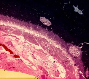
Calcified bone interface, col - red/purple. Mike Kayser. Attribution 4.0 International (CC BY 4.0). Source: Wellcome Collection.

Selected images from this work

View 1 image

About this work

Description

A transmission electron micrograph showing the layers of cells at the interface of calcified bone. The layer of cells across the middle are the osteoblasts. These cells are packed with endoplasmic reticulam (indicating protein synthesis) and are laying down the osteoid - uncalcified collagen matrix (above the osteoblasts). To the right is a developing osteocyte with a mature osteocyte visible in the top centre. The black material is calcified bone.

Contributors

Permanent link