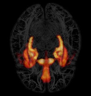
False-colour positron emission tomography (PET) scan highlighting limbic areas in a human brain. A receptor for the inhibitory neurotransmitter GABA is visualised here in regions of the brain associated with memory, learning, emotional processing and addiction, where levels are high. Colour brightness is proportional to signal strength, with the darkest red representing low receptor density, through to bright yellow where the signal is highest. This has been superimposed on a transparent structural representation of the brain. In this orientation, the viewer is looking down at the top of the head, with the eyes facing the bottom edge of the image. To create this image, the receptor was labelled with a radioligand (radioactively labelled chemical that binds to a protein of interest) to enable its detection. 5 individual PET images were taken and then rendered in 3D to give the unique signature set of structures seen here. These include the amygdala, ventral striatum (two central hot spots), the anterior cingulate gyrus (central conical shape), the hippocampi (curved structures on either side) and parts of the temporal lobe. The function of various parts of the limbic system is to regulate impulsivity and response to emotions, such as fear and desire. Visualising neurotransmitters and their receptors gives a unique functional perspective on the brain and allows correlation of function with structures and networks within the brain. This is at the forefront of neuroscientific research in living humans. Horizontal width of image is approximately 18 cm.