On the artificial production of tubercle in the lower animals : a lecture delivered at the Royal College of Physicians, May 15, 1868 / by Wilson Fox.
- Fox, Wilson.
- Date:
- 1868
Licence: Public Domain Mark
Credit: On the artificial production of tubercle in the lower animals : a lecture delivered at the Royal College of Physicians, May 15, 1868 / by Wilson Fox. Source: Wellcome Collection.
11/50
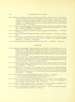
![DESCEIPTION OF PLATES. PLA^rE I. Fk;. 1.—I.ungs of Guinea-pig, containing tubercles, gvoy and semi-transparent at the margins, and in some places slightly 02ia(pie in their centres. ¥\c.. '2.—Enlarged Spleen of Guinea-pig, showing scattered grey granulations, which in some places are agglomerated into groups; in other parts, groups of granulations are seen of oparpie yellow colour. Fk;. :;.—Portion of another Spleeir when the granulations are larger and more scattered. Fio. 4.—Liver of Guinea-pig, shoAving large tracts infiltrated with grey granulations, passing in many 23laces into a more opaf|ue yellow condition. Larger and more isolated ojjai^ue whitish spots are also seen scattered through the tissue. Fig. ;).—Axillary Lymphatics of Guinea-pig, showing cheesy spots. Fig. G.—Subcutaneous Granulations and Cheesy Masses near seat of injury, in rabbit inoculated with tubercle. (These were identical in appearance with those described in the guinea-pig.) The masses are .seen to be composed of agglomerated granulations. Smaller groups of these ai'e seen at variable distances from the larger masses. These latter are greyer and less cheesy than the larger masses. ]'iG. 7.—A Cord of Indurated Tissue, partty cheesy, extending between a lymphatic gland and a chee.sy granulation. (Guinea-pig.) Fit;. 8.—Lobular Pneumonia in a Pabbit, the subject of Laryngo-tracheitis. The infiltration of the pulmonary air-vesicles forms a marked contrast to the granulations in the rabbit. Figs. !), 10.—Lungs of Rabbit with Pyjemic Spots, contrasting with the granulations in the guinea-pig. PLATE IL Fir;. 1. ^I, B.—Tubercular Growth in Sheath of Bronchi. (Guinea-pig.) A X 4 GO diam. shows (a a a) section of bronchial tube at point of bifurcation. The upper part is marked by elastic fibres ; the lower, by cartilage cells, b b h represents the growth of tubercle in its sheath, which is seen to be proceeding by a multiiilication of cells and nuclei, partly round, partly ovate and fusiform. These, at a little distance, are pas.sing into the walls of the alveoli, which are thickened by the growth; the outlines of the alveoli being still maintained. A few enlarged epithelial cells are seen within the alveoli. The vessels of the alveoli so implicated are for the most part obliterated. U X 700 diam. From lower part of A. (a) Sheath of bronchus, (b) Growth of tubercle by round and ovoid cells, (c) Epithelium enlarged and separating. ((7) A growth of fusiform cells which also are seen passing in strings and rows between the capillaries ; of which a good example is observed at (e). Fii;. i'. — GroAvth of Tuliercle in Perivascular Sheath of Pulmonaiy Artery. x 4G0 diam. (reduced). ((iuinea-pig.) (a a). An artery at a point of bifurcation, the section being carried obliquely through the ])lane of both branches. (/' b). ^Multiplication of cells in the .sheath, external to the muscular coat. In both branches a dense agglomeration of these cells is seen in some parts, marked in one Tiranch by (c). The growth is seen extending into the walls of the surrounding air-vesicles, the capillaries of which are impervious to injection. I'l). Enlarged and pigmented epithelial cell.](https://iiif.wellcomecollection.org/image/b20393246_0011.jp2/full/800%2C/0/default.jpg)