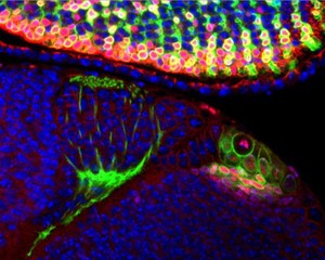
This confocal micrograph shows part of the visual system in a the larval brain of the fruitfly (Drosophila melanogaster). To the right in green are neural stem cells that divide asymmetrically and generate the neurons for the visual system in the brain. These neurons make connections with ingrowing photoreceptor cells (green axons) that derive from the developing eye disc (top).
Using the fruitfly as a model this research assists in understanding how neural stem cells proliferate and divide to generate the large variety of cells that are present in the brain. Studying the switch from symmetric to asymmetric neural stem cell divisions is important in understanding the development of the brain. Symmetric divisions serve to rapidly expand a pool of stem cells whereas asymmetric divisions are required to produce specialised cell types.
mCD8-GFP (green) and Histone2B-mRFP (red) are expressed in the optic lobe neuroblasts (bigger cells to the right) and a subset of photoreceptor cells in the eye disc (top of image). mCD8-GFP (green) also localises to the membrane and thus outlines cells and axons in green. Histone2B-mRFP (red) also labels chromatin in the nuclus. The co-localisation of DAPI (blue) and Histone2B-mRFP (red) results in pink staining.