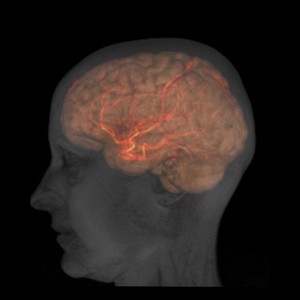
Merged images from two types of MRI scan depicting the head and brain of a healthy female adult. High resolution images of tissue structures in the head and brain were obtained with an anatomical MRI scan. The brain has been segmented and then overlaid with a different colour to differentiate it from the rest of the head.
Magnetic resonance angiography scanning was used to image the large arteries in the brain and provide functional information about the tissue. No injection of contrast agent was required. Contrast in the images was tailored so that inflowing blood appears much brighter than the surrounding tissue. This image was then segmented so that only the brighter blood remains. Using this technique images of the large arteries or veins can be created, which can be useful when investigating pathologies that interrupt the blood supply to the brain, such as a stroke.