Zebrafish embryo

  • Kate Turner & Tom Hawkins
  • Digital Images
  • Online

Available online

view Zebrafish embryo

Attribution 4.0 International (CC BY 4.0)

You can use this work for any purpose, including commercial uses, without restriction under copyright law. You should also provide attribution to the original work, source and licence. Read more about this licence.

Credit

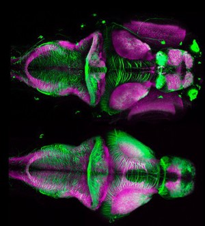
Zebrafish embryo. Kate Turner & Tom Hawkins. Attribution 4.0 International (CC BY 4.0). Source: Wellcome Collection.

Selected images from this work

View 1 image

About this work

Description

A composition showing dorsal views of an undissected and dissected 4 days post fertilisation zebrafish brain. These embryos have been labelled with anti-acetylated tubulin antibody(green) that labels neuronal tracts and anti-SV2(magenta) that stains neuropil. Neuropil is a region between neuronal cell bodies in the gray matter of the brain and blood-brain barrier (i.e. the central nervous system). It consists of a dense tangle of axon terminals, dendrites and glial cell processes. It is where synaptic connections are formed between branches of axons and dendrites.

Permanent link