Fluorescent cranial nerves

  • Dr Jonathan Clarke
  • Digital Images
  • Online

Available online

view Fluorescent cranial nerves

Attribution 4.0 International (CC BY 4.0)

You can use this work for any purpose, including commercial uses, without restriction under copyright law. You should also provide attribution to the original work, source and licence. Read more about this licence.

Credit

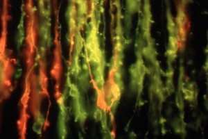
Fluorescent cranial nerves. Dr Jonathan Clarke. Attribution 4.0 International (CC BY 4.0). Source: Wellcome Collection.

Selected images from this work

View 1 image

About this work

Description

A fluorescence micrograph showing nerve cells in the lower part of the chick embryo brain (hindbrain) which have been stained with two differently coloured fluorescent dyes (DiI and DiA). At this stage of development, the two distinct groups of neurons are mixed together, but will later separate to form two cranial nerves, the glossopharyngeal (red) and vagus (green). Biomedical Image Awards 1998.

Contributors

Permanent link