Skip to main content
Wellcome Collection homepage
Visit us
What’s on
Stories
Collections
Get involved
About us
Sign in to your library account
Search for anything
Library account
Search for anything
Search
Images search
Search for images
Search
All
All
Catalogue
Catalogue
Images
Images
Events
Events
Stories
Stories
Colours
Licences
Creative Commons CC-BY (48)
Creative Commons CC-BY-NC (46)
Creative Commons CC0 (1)
Dates
From
to
Types/Techniques
Subjects
Pharmaceutical Preparations (4,000)
Drug Industry (3,967)
London (England) (3,733)
AIDS (Disease) (3,019)
Condoms (2,841)
Acquired Immunodeficiency Syndrome (2,557)
HIV Seropositivity (2,513)
Safe Sex (2,215)
Human anatomy (1,919)
HIV Infections - prevention & control (1,754)
Acquired Immunodeficiency Syndrome - prevention & control (1,701)
AIDS (Disease) - Prevention (1,551)
Great Britain (1,474)
Safe sex in AIDS prevention (1,345)
HIV Seropositivity - transmission (1,275)
Hospitals (1,273)
Death (1,256)
ROYAL VETERINARY COLLEGE (1,107)
Paris (France) (1,079)
Charities (1,054)
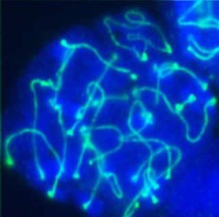
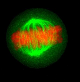
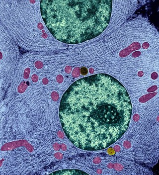
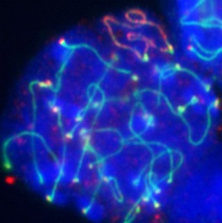
Nucleus (95)
Contributors
Matthew Daniels (33)
NIMR, Francis Crick Institute (16)
University of Edinburgh (14)
Dr Jeremy Skepper (7)
Royal Veterinary College (6)
Kevin Mackenzie, University of Aberdeen (5)
S. Roy & C. Wolff (4)
Anne Weston, Francis Crick Institute (2)
Ezequiel Miron, University of Oxford (2)
Steve Winder (2)
Annie Cavanagh (1)
Christin Bauer (1)
Ludovic Collin (1)
William R. Geddie (1)
Submit
Active filters:
remove
Nucleus
remove
Reset filters
95 results
filtered with: Nucleus
Search result sorting
Sort by:
Relevance
Production dates
Sort order:
Ascending
Descending
Submit
Page
1
of 4
Next (page 2)
Close modal window
Page
1
of 4
Next (page 2)