Wellcome uses cookies.
Read our policy
Close cookie notification
Skip to main content
Wellcome Collection homepage
Visit us
What’s on
Stories
Collections
Get involved
About us
Sign in to your library account
Search our stories, images, catalogue and events
Library account
Search our stories, images, catalogue and events
Search
Images search
Search for images
Search
All
Stories
Images
Catalogue
Events
Colours
Licences
Creative Commons CC-BY (25)
Public Domain Mark (20)
Creative Commons CC-BY-NC (12)
Creative Commons CC0 (3)
In copyright (3)
Creative Commons CC-BY-NC-ND (1)
Dates
From
to
Types/Techniques
Drawings (6)
Engravings (6)
Lithographs (5)
Ink drawings (4)
Oil paintings (4)
Paintings (4)
Photographs (3)
Watercolors (3)
Albumen prints (2)
Book (1)
Catalogue (1)
Photomechanical prints (1)
Prints (1)
Subjects
Vertebrate (16)
Fluorescent probe (14)
Cancer (12)
Amphibia (9)
Aurora B (9)
Cell (9)
Cell culture (9)
Chromosome (9)
DAPI staining (9)
Frog (9)
Human (9)
Blue (8)
Human anatomy (7)
Muscle (6)
Nerves (6)
Vertebrae (6)
Amphibians - anatomy & histology (5)
Anatomy, Comparative (5)
Anatomy, Veterinary (5)
Birds - anatomy & histology (5)
Contributors
Paul Andrews/Univ. Dundee (9)
Dr Guy Bewick, Aberdeen Univ (5)
Retzius, Gustaf, 1842-1919 (5)
Gautier Dagoty, 1717-1785 (4)
Owen, Richard, 1804-1892 (4)
Michael Frank, Royal College of Surgeons (3)
Albinus, Bernhard Siegfried, 1697-1770 (2)
Albinus, Bernhard Siegfried, 1697-1770. Tabulae sceleti et musculorum corporis humani. English (2)
Eustachi, Bartolomeo, -1574 (2)
Fairland, William T., 1805- (2)
Grignion, Charles, 1721-1810 (2)
Royal Veterinary College (2)
Saṅs-rgyas-rgya-mtsho, Sde-srid, 1653-1705 (2)
Scheuchzer, Johann Jakob, 1672-1733 (2)
Schuster, Arthur, Sir, 1851-1934 (2)
Sue, M. (Jean Joseph), 1710-1792 (2)
Thiroux d'Arconville, Marie-Geneviève-Charlotte Darlus, 1720-1805 (2)
Walsh, J (2)
William R. Geddie (2)
Wilson, Erasmus, Sir, 1809-1884 (2)
Submit
82 results
Search result sorting
Sort by:
Relevance
Production dates
Sort order:
Ascending
Descending
Submit
Page
1
of 3
Next (page 2)
Close modal window
Page
1
of 3
Next (page 2)

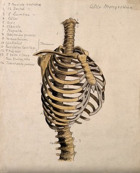
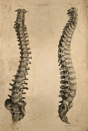
![On the archetype and homologies of the vertebrate skeleton / [Richard Owen].](https://iiif.wellcomecollection.org/image/L0029110/full/600%2C/0/default.jpg)
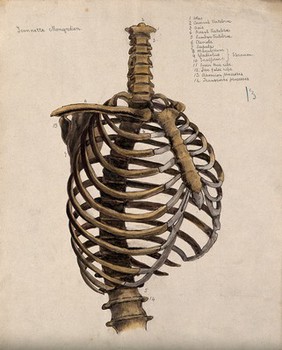
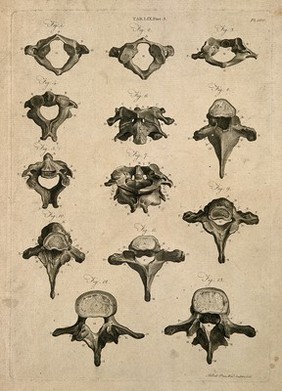
![On the archetype and homologies of the vertebrate skeleton / [Richard Owen].](https://iiif.wellcomecollection.org/image/L0029109/full/420%2C/0/default.jpg)
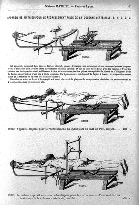
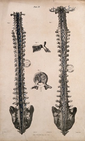
![On the archetype and homologies of the vertebrate skeleton / [Richard Owen].](https://iiif.wellcomecollection.org/image/L0029111/full/420%2C/0/default.jpg)
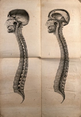
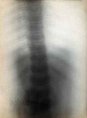
![On the archetype and homologies of the vertebrate skeleton / [Richard Owen].](https://iiif.wellcomecollection.org/image/L0029112/full/600%2C/0/default.jpg)