Wellcome uses cookies.
Read our policy
Close cookie notification
Skip to main content
Wellcome Collection homepage
Visit us
What’s on
Stories
Collections
Get involved
About us
Sign in to your library account
Search our stories, images, catalogue and events
Library account
Search our stories, images, catalogue and events
Search
Images search
Search for images
Search
All
Stories
Images
Catalogue
Events
Colours
Licences
Public Domain Mark (101)
Creative Commons CC-BY (31)
Creative Commons CC-BY-NC (5)
In copyright (3)
Dates
From
to
Types/Techniques
Engravings (56)
Portrait prints (17)
Oil paintings (13)
Allegorical prints (12)
Paintings (12)
Altarpieces (10)
Book illustrations (10)
Woodcuts (9)
Etchings (8)
Pencil works (5)
Drawings (4)
Intaglio prints (4)
Ink drawings (3)
Memento mori (3)
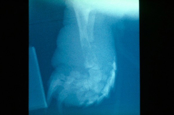
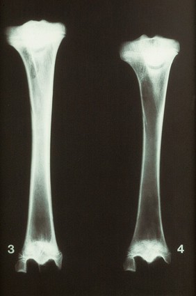
Radiographs (3)
Vanitas (Art) (3)
Lithographs (2)
Photographic prints (2)
Watercolors (2)
Acrostics (1)
Subjects
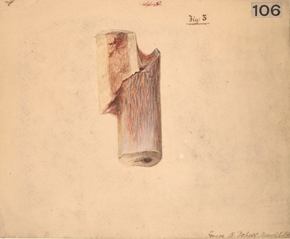
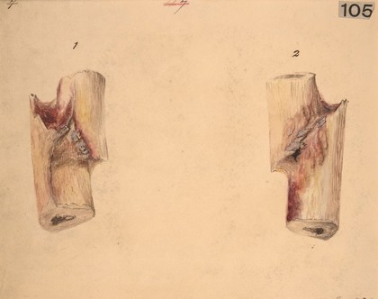
Human anatomy (13)
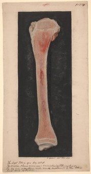
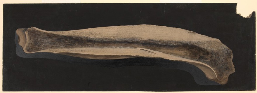
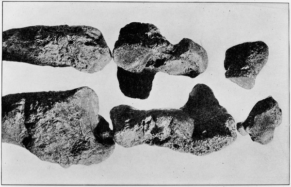
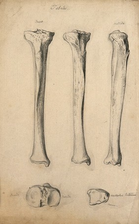
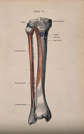
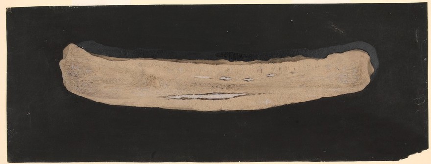
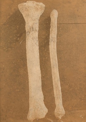
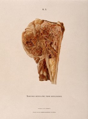
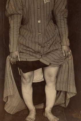
Tibia (12)
Cosmas, Saint (10)
Damian, Saint, -approximately 303 (10)
Dreams (10)
Leg - Transplantation (10)
Surgeons (10)
Surgery, Operative (10)
Transplantation of organs, tissues, etc (10)
3D (9)
Aluminium (9)
Analog (9)
Analogue (9)
Artwork (9)
Black (9)
Circle (9)
Concept (9)
Diagnose (9)
Diagnostics (9)
Digital imaging (9)
Contributors
St Bartholomew's Hospital Archives & Museum (13)
Master of Los Balbases (Painter in Burgos), active approximately 1495 (10)
Sedano, Alonso de (10)
Chiara Salvi (9)
Godart, Thomas (8)
Holbein, Hans, 1497?-1543 (7)
British Library (5)
Cranmer, Thomas, 1489-1556 (5)
Henry Frederick, Prince of Wales, 1594-1612 (5)
Isingrin, M. (Michael), 1500-1557 (5)
Llwyd, Humphrey, 1527-1568 (5)
Lumley, John Lumley, Baron, 1534?-1609 (5)
Manardo, Giovanni, 1462-1536 (5)
Medical Society of London (5)
Petit, John Lewis (5)
Royal Veterinary College (5)
Sims, James, 1741-1820 (5)
Goltzius, Hendrik, 1558-1617 (4)
Wierix, Antonie, -1604 (4)
Vesalius, Andreas, 1514-1564 (3)
Submit
159 results
Search result sorting
Sort by:
Relevance
Production dates
Sort order:
Ascending
Descending
Submit
Page
1
of 6
Next (page 2)
Close modal window
Page
1
of 6
Next (page 2)