Wellcome uses cookies.
Read our policy
Close cookie notification
Skip to main content
Wellcome Collection homepage
Visit us
What’s on
Stories
Collections
Get involved
About us
Sign in to your library account
Search our stories, images, catalogue and events
Library account
Search our stories, images, catalogue and events
Search
Images search
Search for images
Search
All
Stories
Images
Catalogue
Events
Colours
Licences
Creative Commons CC-BY (8)
Dates
From
to
Types/Techniques
Photographs (2)
Portrait photographs (2)
Subjects
Flu (8)
Influenza (8)
Pandemic (8)
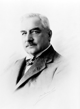
Adami, J. George (John George), 1862-1926 (2)
Contributors
John George Adami (8)
Submit
10 results
Search result sorting
Sort by:
Relevance
Production dates
Sort order:
Ascending
Descending
Submit
Page
1
of 1
Close modal window
Page
1
of 1


![Drawing of the 1918 Influenza: Trachea showing earliest stage of reaction in influenza. Epithelium intact, but basement [?] slightly swollen as [?], with congestion and some small cells influtration of sub[?]. Hyaline deposits over epithelium](https://iiif.wellcomecollection.org/image/L0041257/full/282%2C/0/default.jpg)
![Drawing of the 1918 Influenza: Trachea showing particular retention of epithelium, by, and in the neighbourhood of duet [?] of one of the mucus glands.](https://iiif.wellcomecollection.org/image/L0041258/full/282%2C/0/default.jpg)