Wellcome uses cookies.
Read our policy
Close cookie notification
Skip to main content
Wellcome Collection homepage
Visit us
What’s on
Stories
Collections
Get involved
About us
Sign in to your library account
Search our stories, images, catalogue and events
Library account
Search our stories, images, catalogue and events
Search
Images search
Search for images
Search
All
Stories
Images
Catalogue
Events
Colours
Licences
Creative Commons CC-BY (91)
Creative Commons CC-BY-NC (22)
Creative Commons CC0 (2)
Dates
From
to
Types/Techniques
Subjects
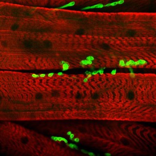
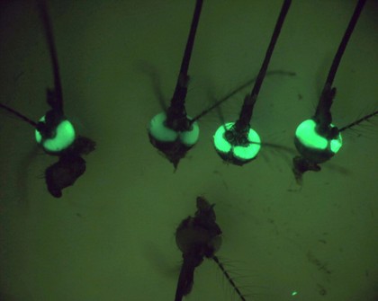
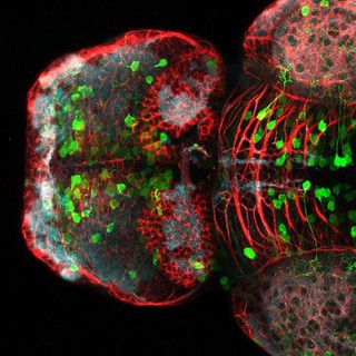
GFP (28)
Cancer (26)
DAPI staining (24)
Model organism (21)
Blue (20)
Green (19)
Cell (18)
Cell culture (18)
Aurora B (17)
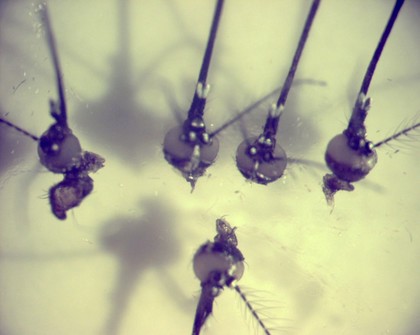
Fruit fly (17)
Human (17)
Transgenic (17)
Chromosome (16)
Fluorescent probe (16)
Green fluorescent protein (16)
Insect (15)
Red (15)
Development (12)
Embryo (11)
Vertebrate (11)
Contributors
Derric Nimmo & Paul Eggleston (16)
Paul Andrews/Univ. Dundee (15)
Alex Gray (8)
Fernan Federici & Jim Haseloff (8)
Paul Appleton, University of Dundee (5)
Fernán Federici (4)
Hermann Aberle, University of Munster (3)
Kate Turner, Dr Steve Wilson (3)
Monica Folgueira & Steve Wilson (3)
Valeria Molinari, Louise Howell, Maria Vinci, Katy Taylor and Chris Jones, Institute of Cancer Research (3)
Yirui Sun (3)
Aamir Ahmed, Jane Pendjiky and Michael Millar (2)
Alison Dun, ESRIC (Edinburgh Super-Resolution Imaging Consortium) (2)
Daniela Malide, Jean-Yves Metais, Cynthia E Dunbar, NIH, Bethesda, USA (2)
Dr Andrea H. Brand (2)
Dr Mónica Folgueira (2)
Dr Steve Wilson (2)
Kate Storey (2)
Prof. Bill Harris (2)
S. Roy & F. Muller (2)
Submit
115 results
Search result sorting
Sort by:
Relevance
Production dates
Sort order:
Ascending
Descending
Submit
Page
1
of 4
Next (page 2)
Close modal window
Page
1
of 4
Next (page 2)