Wellcome uses cookies.
Read our policy
Close cookie notification
Skip to main content
Wellcome Collection homepage
Visit us
What’s on
Stories
Collections
Get involved
About us
Sign in to your library account
Search our stories, images, catalogue and events
Library account
Search our stories, images, catalogue and events
Search
Images search
Search for images
Search
All
Stories
Images
Catalogue
Events
Colours
Licences
Public Domain Mark (24)
Creative Commons CC-BY (23)
Creative Commons CC-BY-NC (3)
In copyright (2)
Dates
From
to
Types/Techniques
Lithographs (11)
Engravings (9)
Anatomical fugitive sheets (8)
Book illustrations (5)
Mezzotints (5)
Oil paintings (3)
Paintings (3)
Collotypes (2)
Drawings (2)
Etchings (2)
Motion study photographs (2)
Prints (2)
Bookplates (1)
Ink drawings (1)
Lantern Slide (1)
Portrait prints (1)
Watercolors (1)
Wood engravings (1)
Subjects
Human anatomy (24)
Muscles (13)
Anatomy (10)
Animal Structures (8)
Eye - Anatomy (7)
Eye - Muscles (7)
Head (7)
Countryside (6)
Garden (6)
Herbal remedies (6)
Petal (6)
Poison (6)
Arteries (4)
Face (4)
Artwork (3)
Blind (3)
Clay (3)
Cranium (3)
Feel (3)
Forensic science (3)
Contributors
Rabaud, Etienne, 1868- (8)
Dr Henry Oakeley (6)
Alembert, Jean Le Rond d', 1717-1783 (4)
Diderot, Denis, 1713-1784 (4)
Haller, Albrecht von, 1708-1777 (4)
Heather Spears (4)
Rollinus, C. J., active 18th century (4)
Ruysch, Frederik, 1638-1731 (4)
Albinus, Bernhard Siegfried, 1697-1770 (3)
Duverney, M. (Jacques-François-Marie), 1661-1748 (3)
Gautier Dagoty, 1717-1785 (3)
Heather Spears, photography ICandy (3)
Lobley, John Hodgson, 1878-1954 (3)
Bénard, 1731-1794 (2)
Bidloo, Govard, 1649-1713 (2)
Ford, G. H. (George Henry) (2)
Gautier d'Agoty, A. E. (Arnaud Eloy), 1741-1771 (2)
Heister, Lorenz, 1683-1758 (2)
Jadelot, Jean-Nicolas, 1738-1793 (2)
University of Pennsylvania (2)
Submit
63 results
Search result sorting
Sort by:
Relevance
Production dates
Sort order:
Ascending
Descending
Submit
Page
1
of 3
Next (page 2)
Close modal window
Page
1
of 3
Next (page 2)


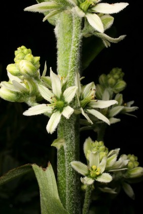
![Atropa belladonna L. Solanaceae. Deadly nightshade. Dwale. Morella, Solatrum, Hound's berries, Uva lupina, Cucubalus, Solanum lethale. Atropa derives from Atropos the oldest of the three Fates of Greek mythology who cut the thread of Life (her sisters Clotho and Lachesis spun and measured the thread, respectively). belladonna, literally, means 'beautiful lady' and was the Italian name for it. Folklore has it that Italian ladies put drops from the plant or the fruits in their eyes to make themselves doe-eyed, myopic and beautiful. However, this is not supported by the 16th and 17th century literature, where no mention is ever made of dilated pupils (or any of the effects of parasympathetic blockade). Tournefort (1719) says 'The Italians named this plant Belladonna, which in their language signifies a beautiful woman, because the ladies use it much in the composition of their Fucus [rouge or deceit or cosmetic] or face paint.' Parkinson says that the Italian ladies use the distilled juice as a fucus '... peradventure [perhaps] to take away their high colour and make them looke paler.' I think it more likely that they absorbed atropine through their skin and were slightly 'stoned' and disinhibited, which made them beautiful ladies in the eyes of Italian males. Distribution: Europe, North Africa, western Asia. Culpeper (1650) writes: 'Solanum. Nightshade: very cold and dry, binding … dangerous given inwardly … outwardly it helps the shingles, St Antonie's Fire [erysipelas] and other hot inflammation.' Most of the 16th, 17th and 18th century herbals recommend it topically for breast cancers. Poisonous plants were regarded as 'cold' plants as an excess of them caused death and the body became cold. They were regarded as opposing the hot humour which kept us warm and alive. Poultices of Belladonna leaves are still recommended for muscle strain in cyclists, by herbalists. Gerard (1633) writes that it: 'causeth sleep, troubleth the mind, bringeth madnesse if a few of the berries be inwardly taken, but if more be taken they also kill...'. He was also aware that the alkaloids could be absorbed through the skin for he notes that a poultice of the leaves applied to the forehead, induces sleep, and relieves headache. The whole plant contains the anticholinergic alkaloid atropine, which blocks the peripheral actions of acetylcholine in the parasympathetic nervous system. Atropine is a racemic mixture of d- and l- hyoscyamine. Atropine, dropped into the eyes, blocks the acetylcholine receptors of the pupil so it no longer constricts on exposure to bright light - so enabling an ophthalmologist to examine the retina with an ophthalmoscope. Atropine speeds up the heart rate, reduces salivation and sweating, reduces gut motility, inhibits the vertigo of sea sickness, and is used to block the acetylcholine receptors to prevent the effects of organophosphorous and other nerve gas poisons. It is still has important uses in medicine. Atropine poisoning takes three or for days to wear off, and the hallucinations experienced by its use are described as unpleasant. We have to be content with 'madness', 'frenzie' and 'idle and vain imaginations' in the early herbals to describe the hallucinations of atropine and related alkaloids as the word 'hallucination' in the sense of a perception for which there is no external stimulus, was not used in English until 1646 (Sir T. Browne, 1646). It is a restricted herbal medicine which can only be sold in premises which are registered pharmacies and by or under the supervision of a pharmacist (UK Medicines and Healthcare Products Regulatory Agency (MHRA)). Photographed in the Medicinal Garden of the Royal College of Physicians, London.](https://iiif.wellcomecollection.org/image/B0008945/full/282%2C/0/default.jpg)